Patient information leaflets
Trusted patient information leaflets with clear, clinically reviewed advice on conditions, symptoms and treatments.
Health topics
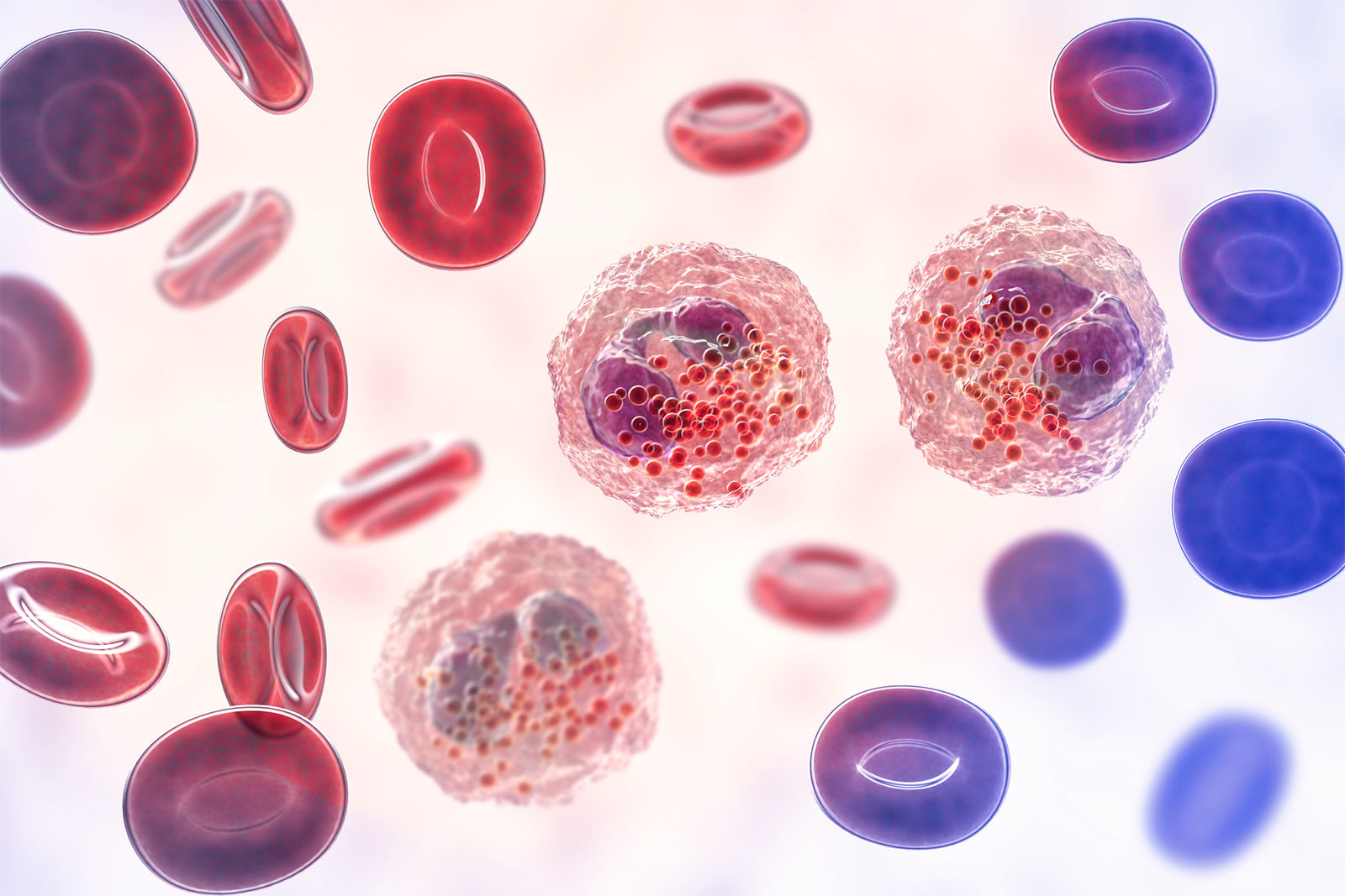
Allergies, blood and immune system
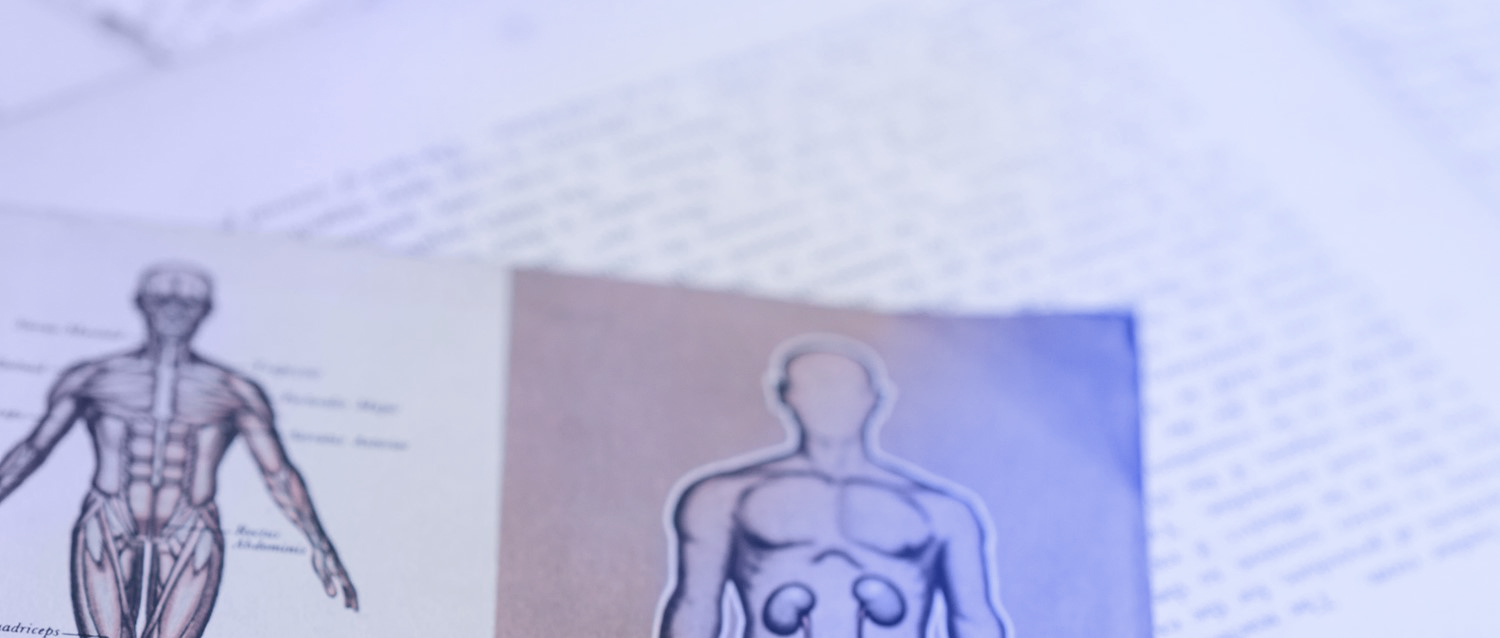
Bones, joints and muscles
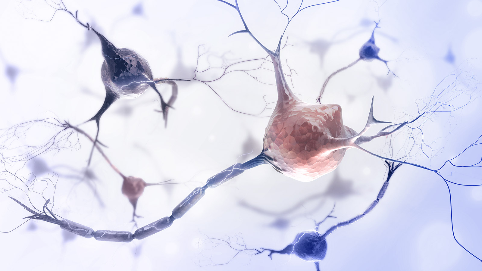
Brain and nerves

Cancer
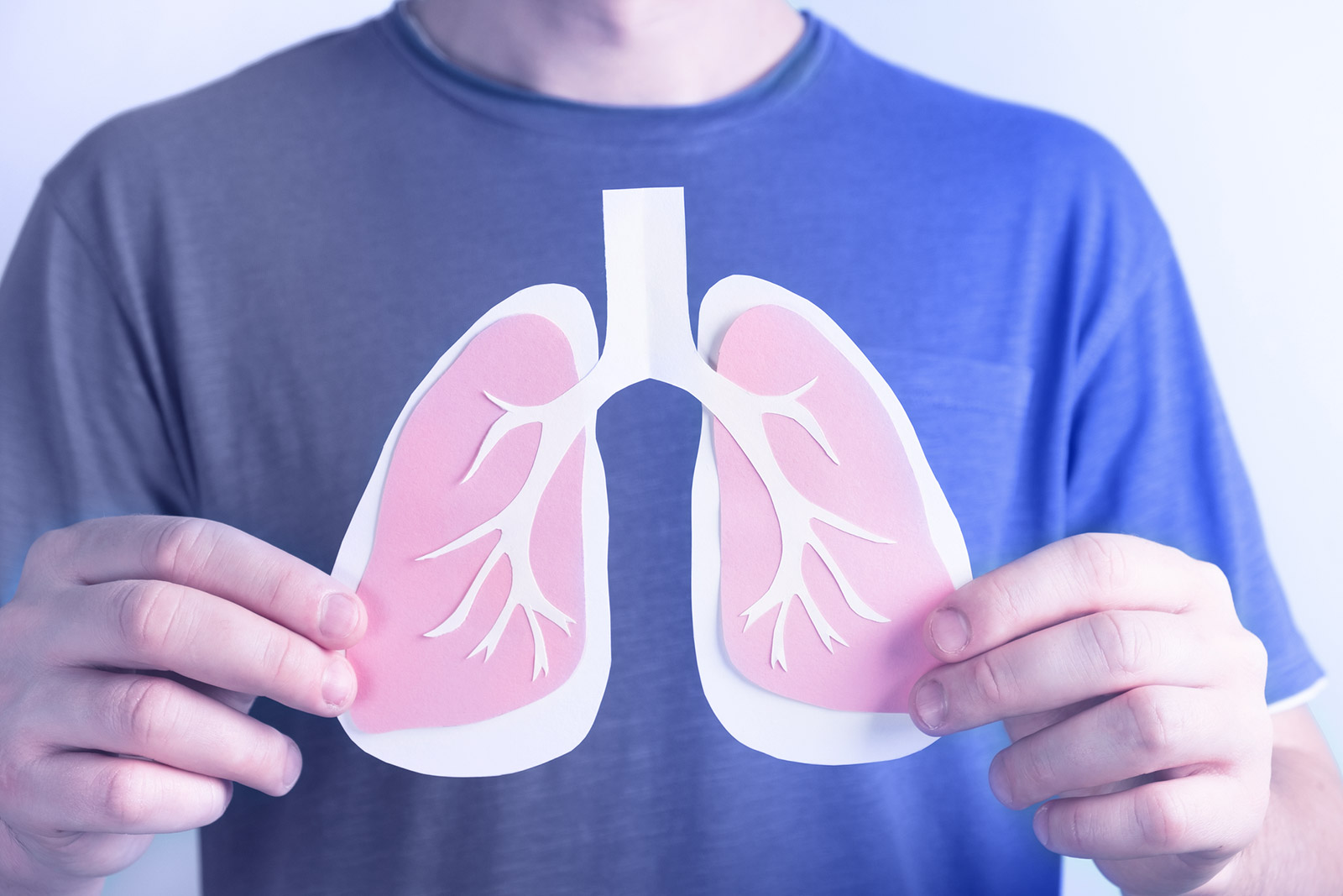
Chest and lungs

Children's health
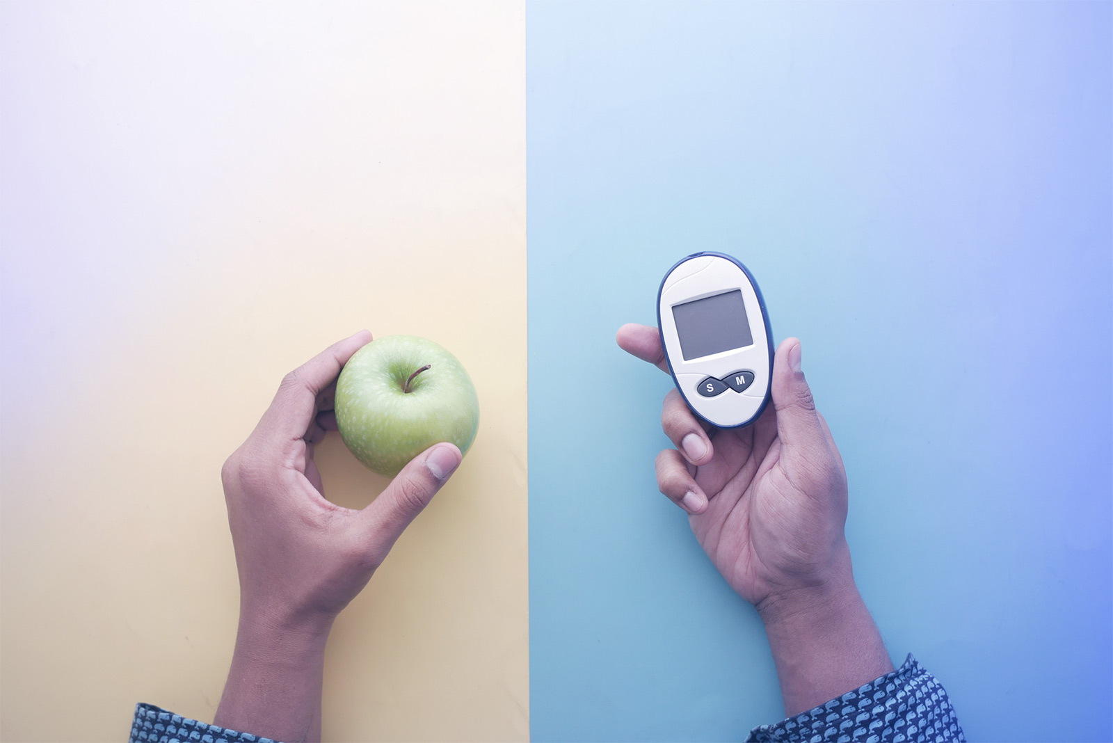
Diabetes

Digestive health

Ear, nose and throat
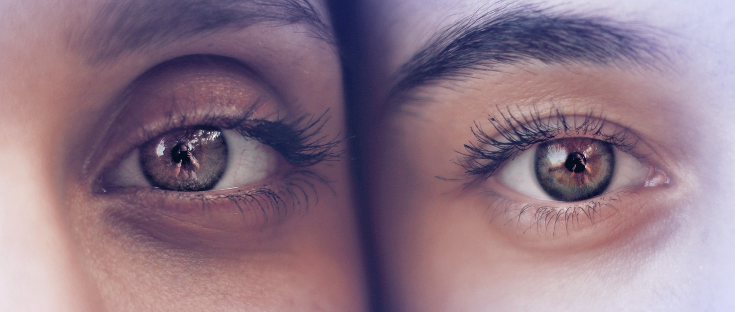
Eye health

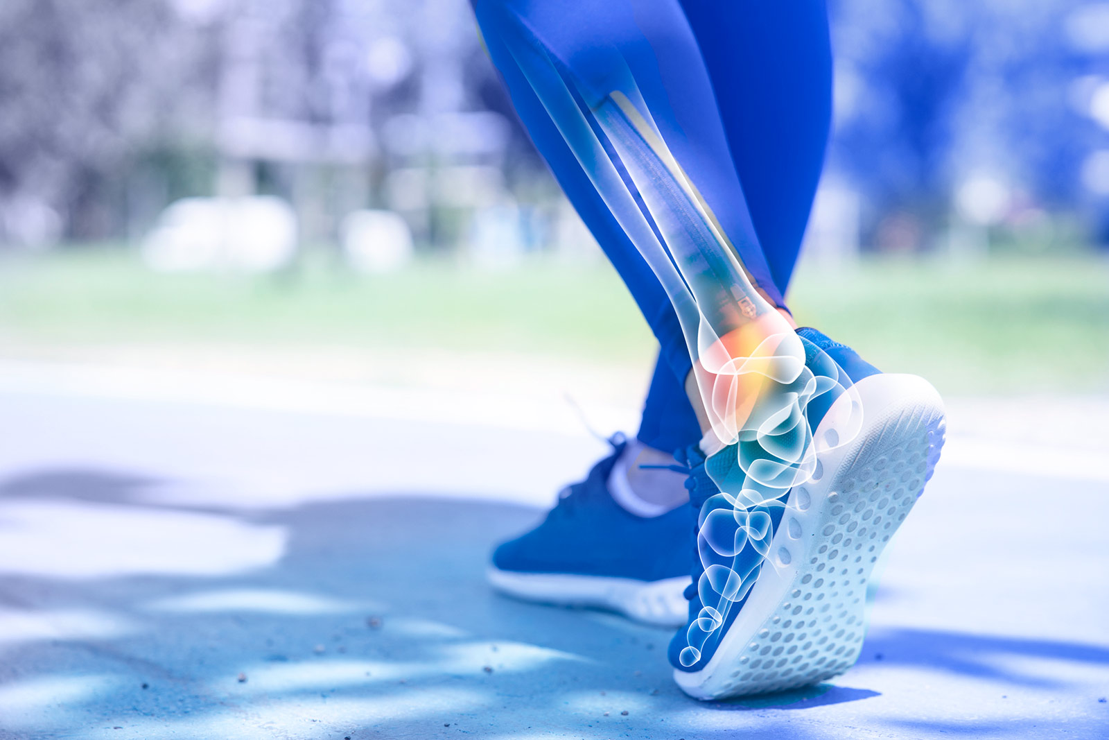
Foot care

General health

Healthy living
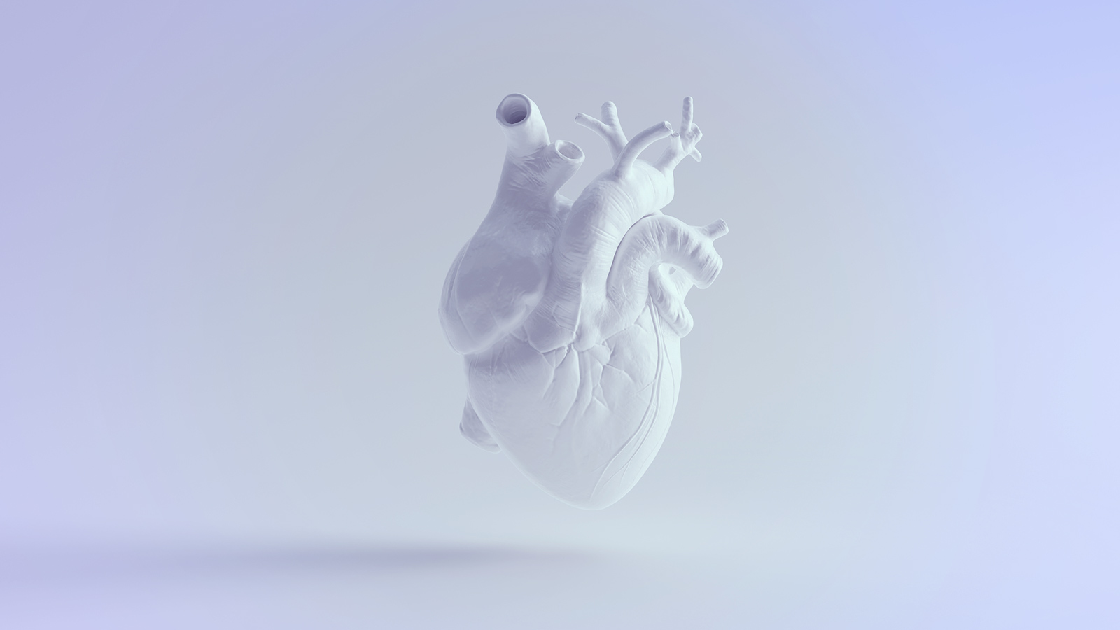
Heart health and blood vessels

Hormones

Infections
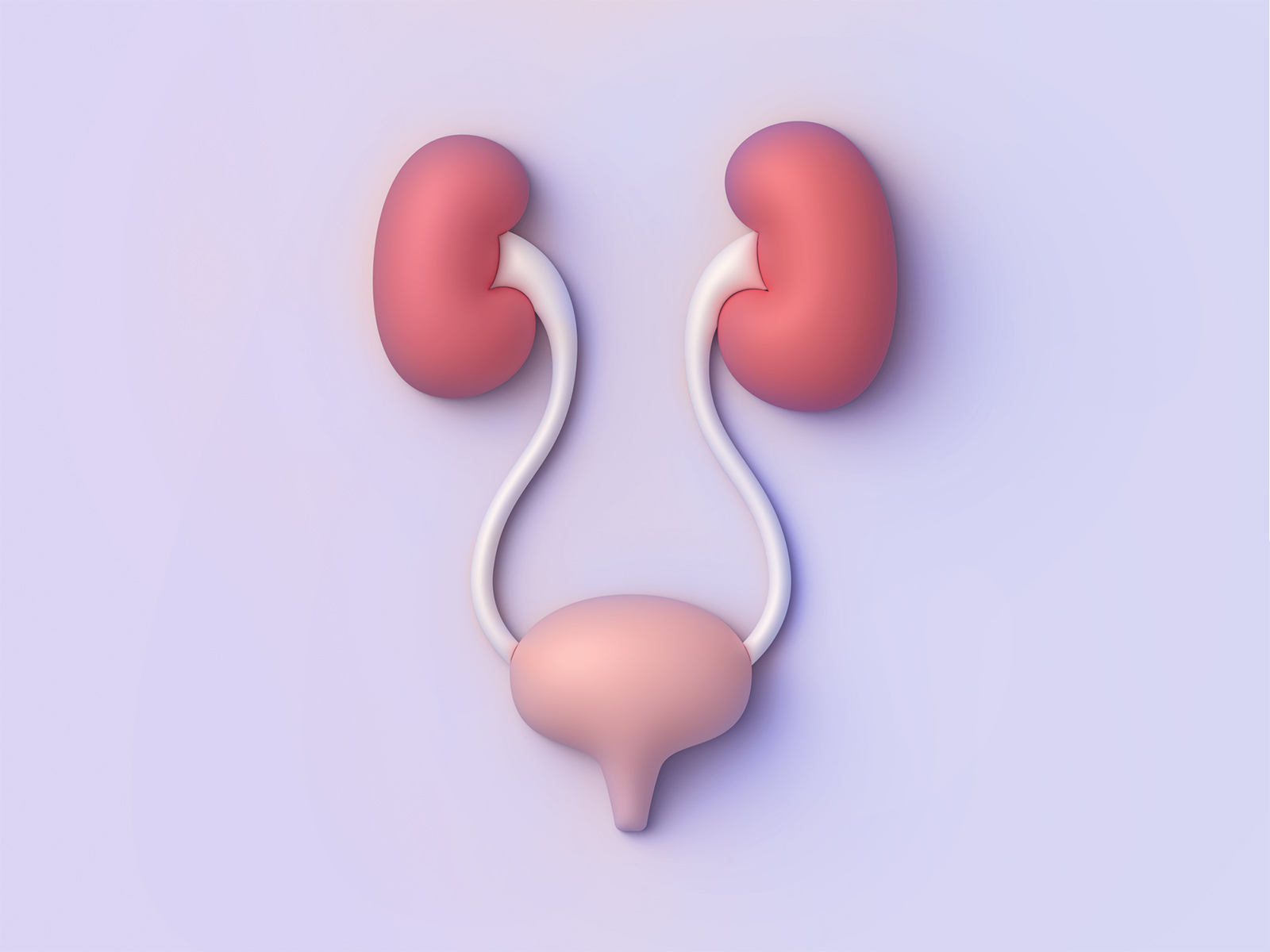
Kidney and urinary tract

Men's health

Mental health

Oral and dental care

Pregnancy

Senior health

Sexual health
Signs and symptoms
Skin, nail and hair health
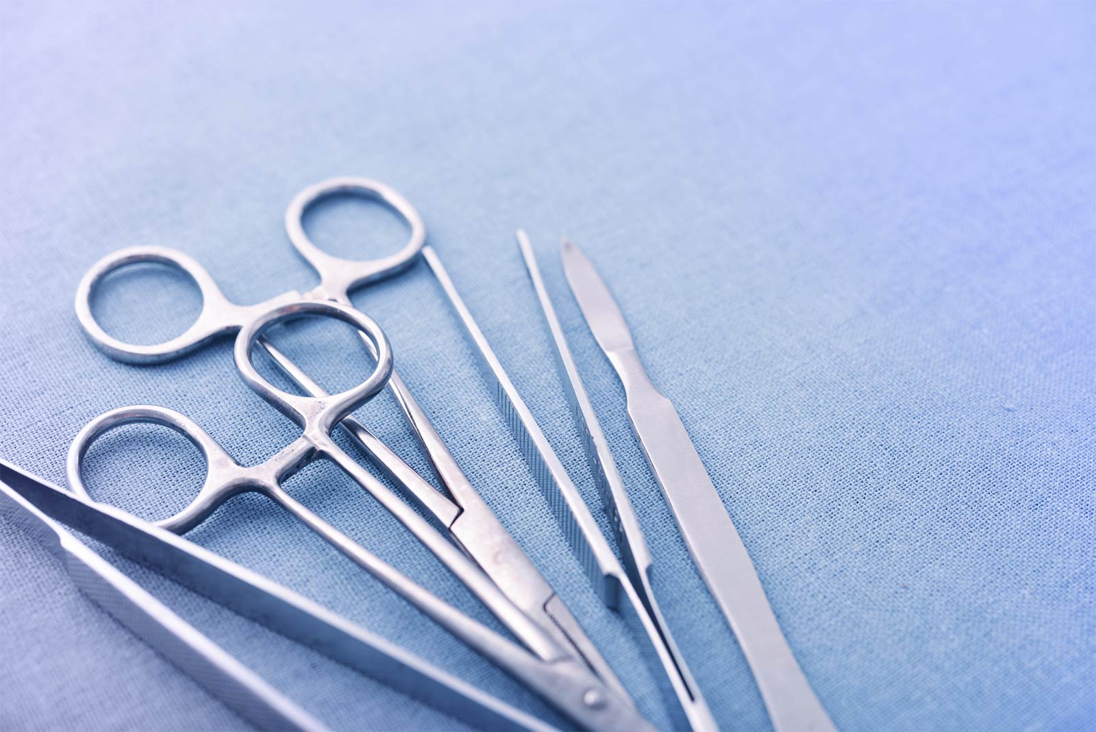
Surgery and procedures
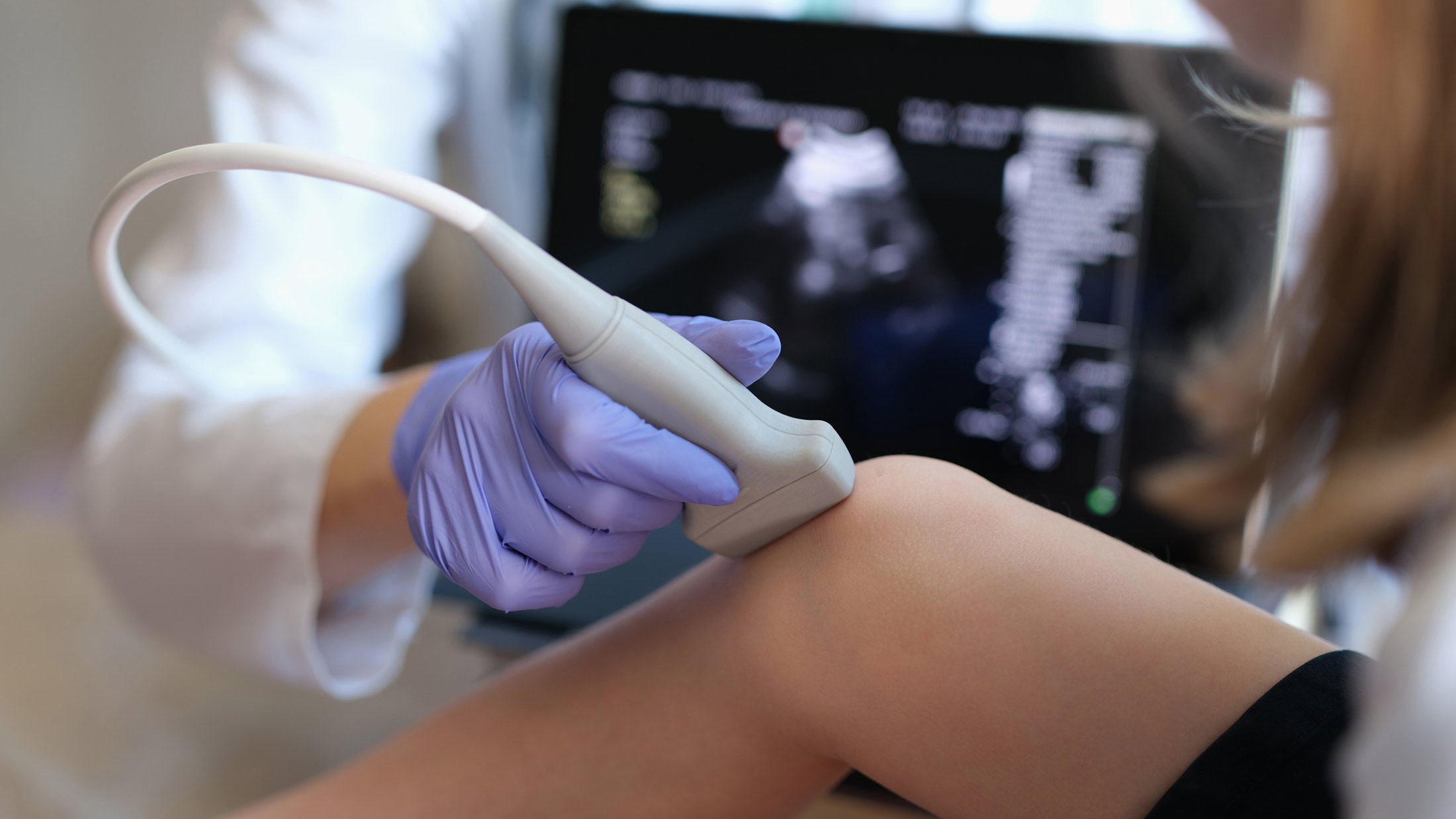
Tests and investigations

Travel and vaccinations

Treatment and medication

Women's health
A
- Abdominal aortic aneurysm
- Abdominal masses
- Abdominal pain
- Abdominal pain - left lower
- Abdominal pain - left upper
- Abdominal pain - right lower
- Abdominal pain - right upper
- Abdominal pain in children - left-sided
- Abdominal pain in children - recurrent
- Abdominal pain in children - right lower
- Abdominal pain in pregnancy - left lower
- Abdominal pain in pregnancy - right lower
- Abdominoplasty
- Abnormal heart rhythms
- Abortion
- Abscess
- Abscess - dental
- Absence seizures
- ACE inhibitors
- Achalasia
- Achilles tendinopathy
- Achilles tendon rupture
- Acid reflux and oesophagitis
- Acid reflux in pregnancy
- Acne
- Acne treatments
- Acoustic neuroma
- Acromegaly
- Actinic keratosis
- Acute angle-closure glaucoma
- Acute bronchitis
- Acute coronary syndrome
- Acute coronary syndrome - heart attack
- Acute diarrhoea in children
- Acute exacerbations of COPD
- Acute kidney injury
- Acute lymphoblastic leukaemia
- Acute myeloid leukaemia
- Acute pancreatitis
- Acute prostatitis
- Acute renal failure
- Acute sinusitis
- Acute stress reaction
- Addison's disease
- ADHD in adults
- ADHD in children
- ADHD self-assessment tool
- Adhesive capsulitis
- Adolescence
- Adrenal fatigue
- Adrenarche
- Adult sepsis safety net
- Aeroplane ear
- Ageing
- Ageing feet
- Agoraphobia
- AIDS - acquired immune deficiency syndrome
- Albinism
- Alcohol and drug support services NHS - refer yourself
- Alcohol and liver disease
- Alcohol and sensible drinking - safe limits of alcohol
- Alcohol units calculator
- Alcohol withdrawal and alcohol detoxification
- Alcoholism and problem drinking
- ALL - acute lymphoblastic leukaemia
- Allergic conjunctivitis
- Allergic rhinitis - seasonal
- Allergies
- Allergy - cow's milk protein
- Allergy - drug
- Allergy - food (and intolerance)
- Allergy - house dust mite and pet
- Allergy - nut
- Allergy - oral allergy syndrome
- Allergy - vaccines containing egg
- Allergy test - skin prick
- Alopecia - androgenetic (male pattern baldness)
- Alopecia areata
- Alpha-1 antitrypsin deficiency
- Alpha-blockers
- ALS - amyotrophic lateral sclerosis
- Alternative medicine
- Alternatives to HRT for menopause symptoms
- Altitude sickness
- Alzheimer's disease
- Amblyopia (lazy eye)
- Ambulatory blood pressure monitoring
- Ambulatory electrocardiogram
- Amenorrhoea
- Aminosalicylates
- AML - acute myeloid leukaemia
- Amniocentesis
- Amoebiasis
- Amphetamines
- Amyl nitrates
- Anaemia
- Anaemia - iron deficiency
- Anaemia - macrocytic
- Anaesthesia
- Anaesthesia - death risk
- Anaesthetic for hip or knee replacement
- Anal disorders - local preparations
- Anal fissure
- Anal fistula
- Anal pain - proctalgia fugax
- Analgesics
- Anaphylaxis
- Aneurysm (abdominal aortic)
- Anger management
- Angina
- Angina - microvascular
- Angina - Prinzmetal's
- Angio-oedema
- Angiography - cerebral
- Angiography - coronary
- Angioplasty - coronary
- Ankle injury
- Ankylosing spondylitis
- Anogenital warts
- Anorexia nervosa
- Anosmia
- Antacids
- Antenatal care NHS - refer yourself
- Antenatal checks
- Anthelmintics
- Anti-inflammatory tablets
- Antibiotics
- Antibiotics, germs and microbes
- Antibody and antigen tests
- Anticoagulants
- Antidepressants
- Antifungal medication
- Antihistamines
- Antiphospholipid syndrome
- Antipsychotic medicines
- Antispasmodics
- Antithyroid medicines
- Anxiety
- Anxiety in children
- Aortic dissection
- Aortic regurgitation
- Aortic stenosis
- Apathy
- Aphasia
- Aphthous mouth ulcers
- Appendicitis
- Appendicitis symptoms
- Appetite loss
- Are e-cigarettes safe?
- Arrhythmias
- Arterial blood gases
- Arthritis
- Arthritis - juvenile
- Arthritis - psoriatic
- Arthritis - reactive
- Arthritis - rheumatoid
- Arthritis - septic
- Arthroscopy and arthroscopic surgery
- AskPatient
- Aspiration pneumonia
- Aspirin and other antiplatelet medicines
- Assisted delivery
- Asthma
- Asthma Control Test (ACT)
- Asthma inhalers
- Asthma peak flow meter
- Asthma- brittle
- Astigmatism
- Atheroma
- Athlete's foot
- Atkins diet
- Atopic eczema
- Atrial fibrillation
- Atrial fibrillation and stroke prevention
- Atrial septal defect
- Atrophic vaginitis
- Audiology
- Audiometry
- Auditory processing disorder
- Autism spectrum disorders
- Autoimmune hepatitis
- Avoidant personality disorder
- Axial spondyloarthritis